Biological Electron Microscopy Facility
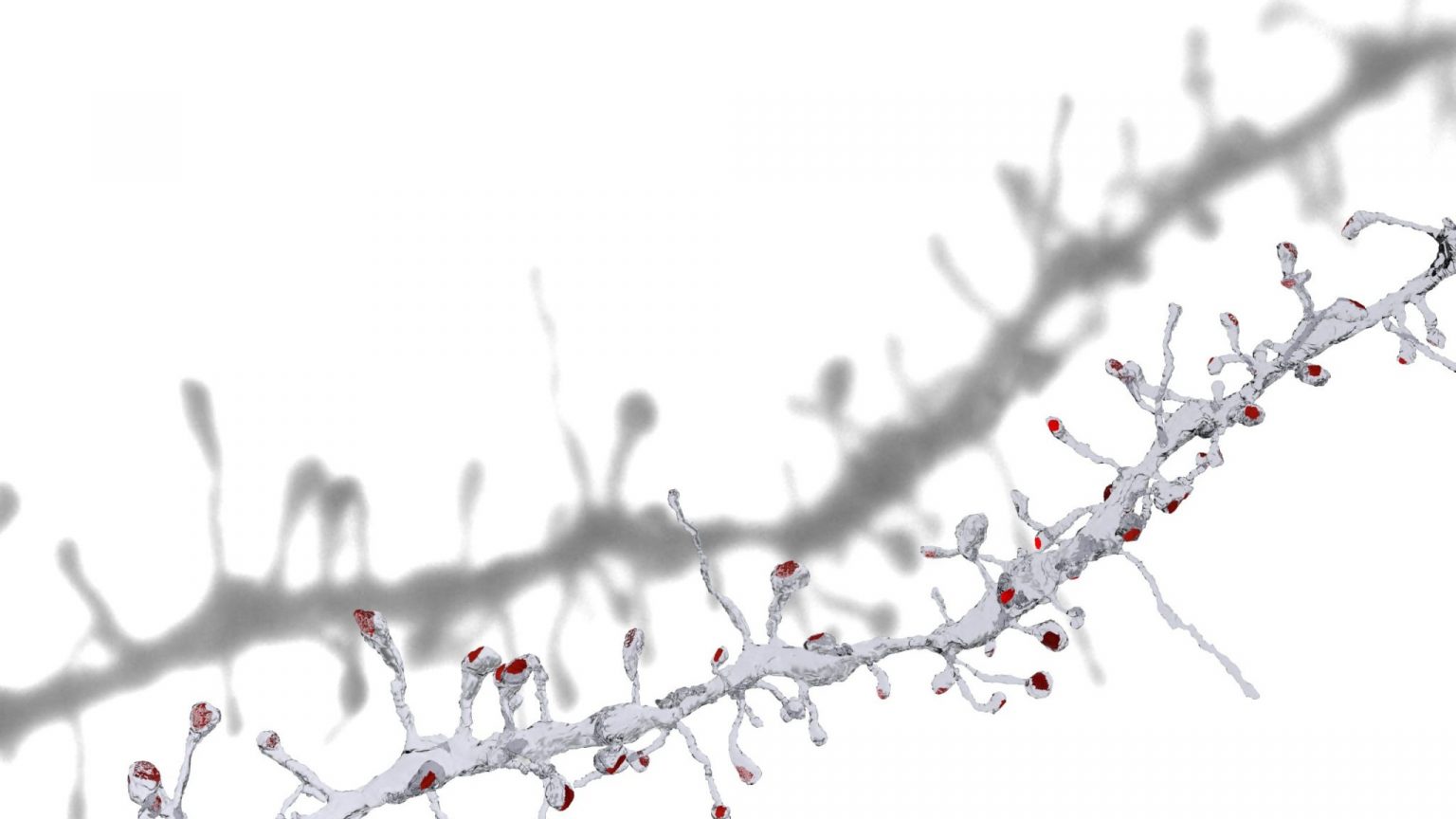
3d reconstruction of a dendrite showing spines and their synapses (in red). This work was done as part of a project carried out for Sami El Boustani (University of Geneva), and published in Science in 2018.
Latest news
Effect of human Tau protein on fly development imaged with SEM
A recent study by the group of Brain McCabe uses genetic, microscopy and computational methods to assess the effect of the human tau protein on the development of individual neurons. This protein, involved in Alzheimer’s disease, when expressed in individual neurons resulted in significant structural and functional deficits within the cell. More explanations can be seen here, and details of the project can be found here.
Role of perivascular cells in the gut revealed by large-scale 3D EM
In a collaboration with the group of Tatiana Petrova (Department of Oncology, UNIL; ISREC, EPFL) the BioEM Facility has carried out correlative light and 3D electron microscopy on samples of mouse gut to investigate the organisation and architecture of telocytes – cells that surround blood capillaries. Block face scanning electron microscopy, at an unprecedented scale, in the region of the villus tip, revealed that the telocytes are arranged in such a way that they favour the availability of particular growth factors to therefore influence blood vessel patterning. The study can be viewed here.
Block face scanning EM in bladder infection
The group of John McKinney used the Block face scanning EM technique to visualize the infection of a bladder with Uropathogenic Escherichia coli (UPEC). This technique reveals that solitary bacteria can be found throughout the bladder wall and may be intracellular or pericellular (sandwiched between uroepithelial cells). The study can be viewed here.
Movie: 3D reconstruction of the infected bladder
TEM and Block face scanning EM to analyse the effect of synaptic develpment in absence of regulated vesicular release.
The group of Zoltan Molnar from the university of Oxford explore the effect of the absence of regulated vesicular release on the synapse morphogenesis and maturation in regions that receive large and powerful cortical inputs such as in the thalamus. This question was explored using 3D electron microscopy and immunoelectron microscopy analyses in the large, complex synpases formed between cortical sensory efferent axons and dendrites in the posterior thalamic nucleus. the study can be viewed here.
Graham Knott, office AI0143
tel: +41 21 6931862